|
Érythroplasie
Érythroplasie. Noter la lésion rouge et irrégulière dans la muqueuse de la joue, à côté des zones blanches.
|
|
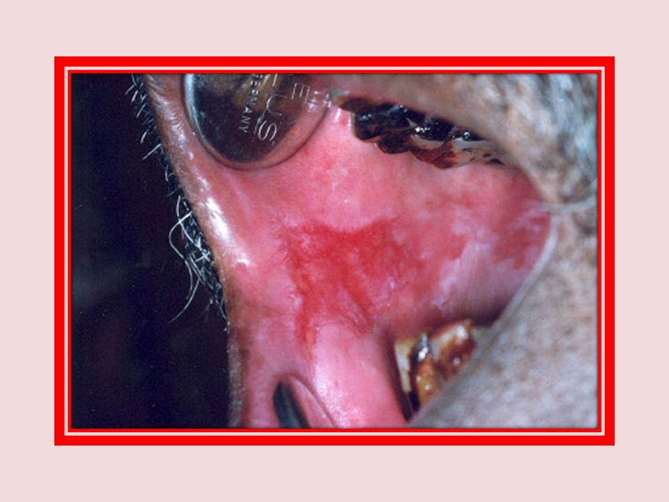
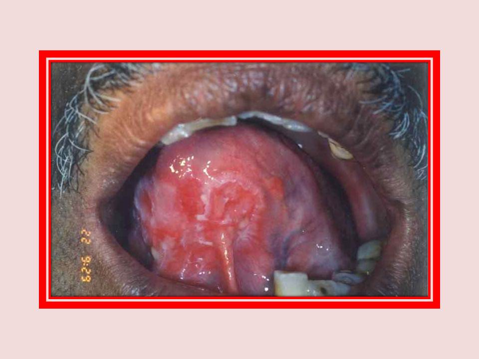
Érythroplasie
Érythroplasie. Notez la lésion rouge, irrégulière, velouté sur le côté droit de la voûte palatine. Il est cliniquement suspectée de transformation maligne.
|
|
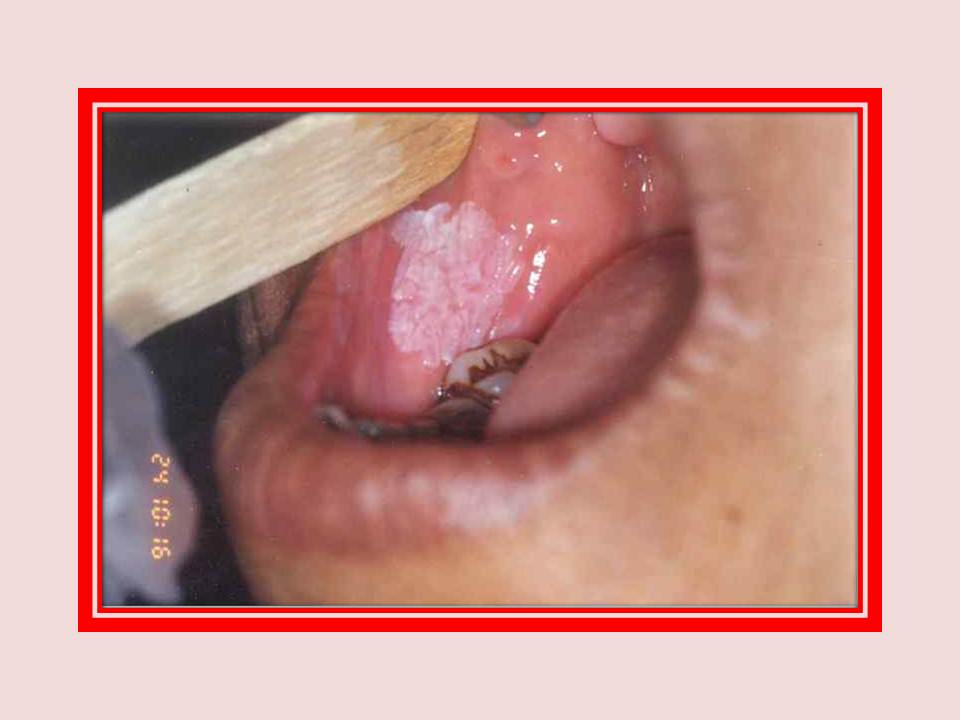
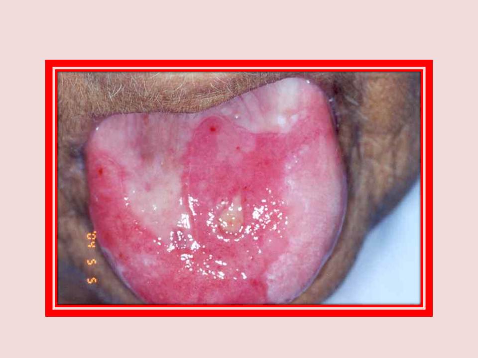
Leucoplasie
Leucoplasie homogène muqueuse de la joue.
|
|
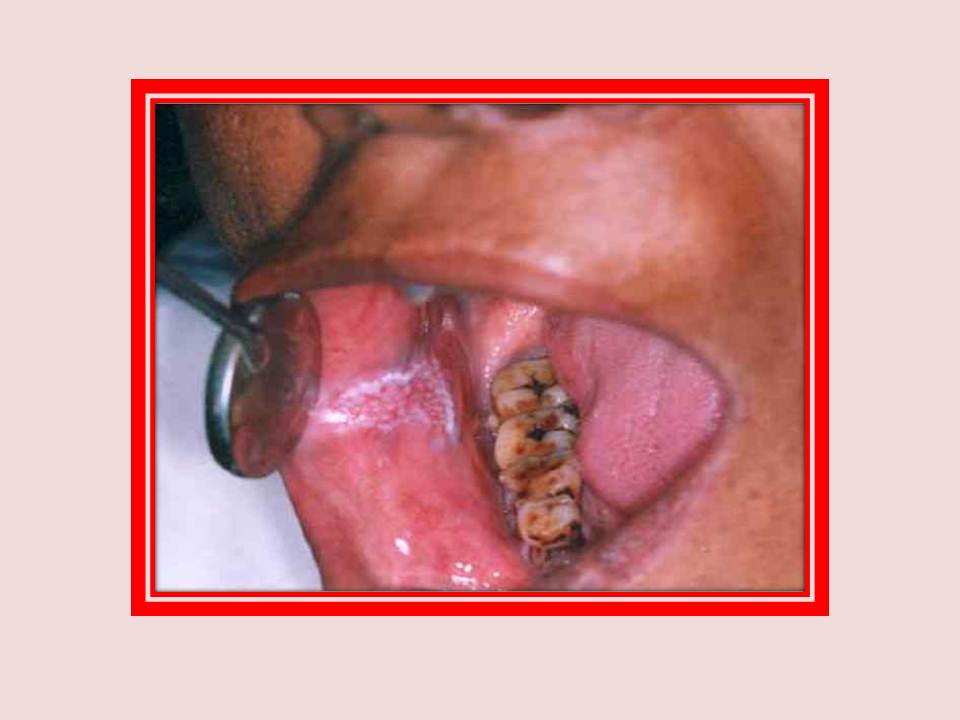
Leucoplasie
Leucoplasie nodulaire. Une lésion bien circonscrite de 3x1 cm de la muqueuse de la joue. Notez les points comme des têtes d'épingle éparpillés sur une base érythémateuse.
|
|
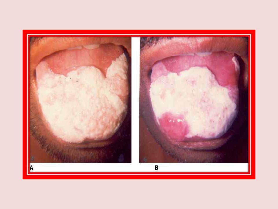
Leucoplasie
Figure A: candidose chez un patient présentant une leucoplasie homogène.
Figure B: Le même patient trois semaines après le traitement antifongique. La candidose régresse et la leucoplasie reste.
|
|
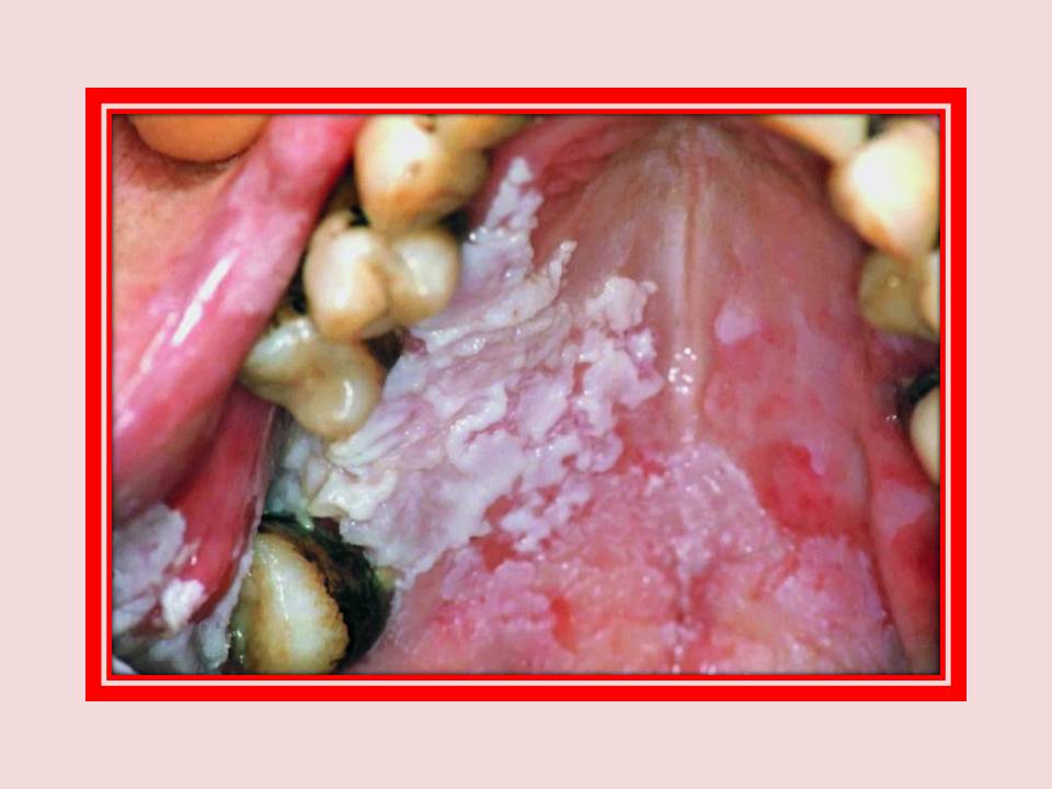
Leucoplasie
Leucoplasie proliférative verruqueux (PVL). Notez les plaques blanches, larges, épaisses et adhérentes.
|
|
Lichen Planus
Lichen Planus. Notez la couleur blanchâtre de 4x3.5 cm lésion sur le côté droit de la langue entrecoupées de zones de pigmentation. Le lichen plan annulaire peut être vu sur le côté gauche de la face dorsale de la langue.
|
|
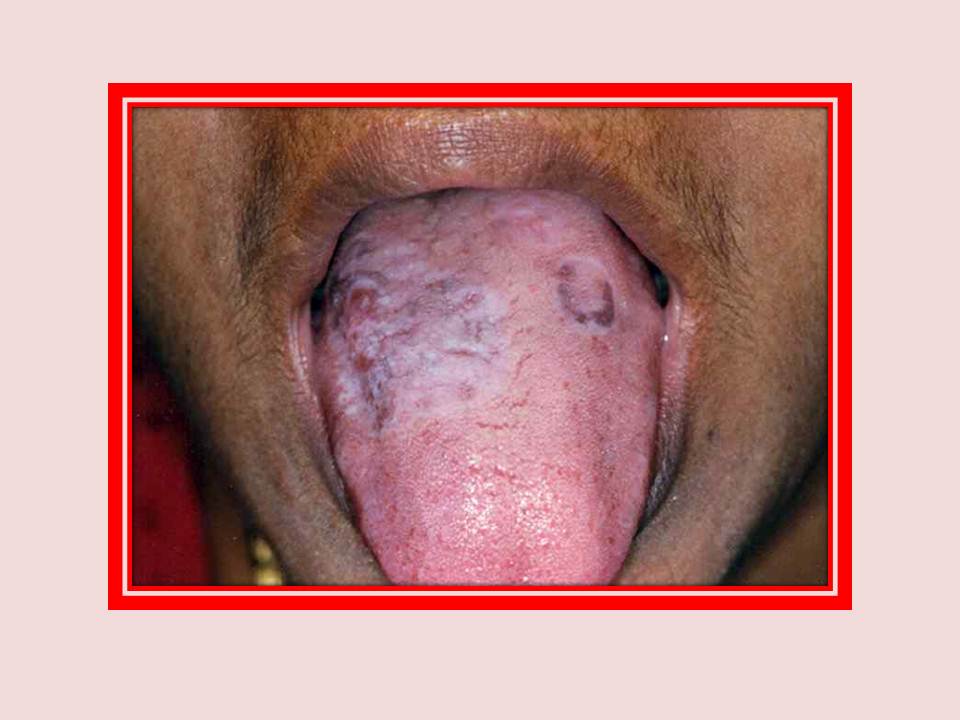
Lichen Planus
Lichen plan annulaire. Notez le modèle des formes similaires à des anneaux.
|
|
Lichen Planus
Lichen plan érosif. Les zones érodées entourées par des rainures similaires à des anneaux.
|
|
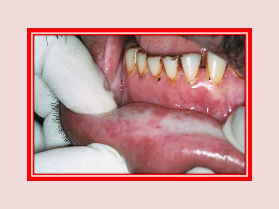
Fibrose sous-muqueuse orale
Noter la zone blanchâtre au fond de la muqueuse labiale.
|
|
Fibrose sous-muqueuse orale
fibrose sous-muqueuse orale avec érythroplasie.
|
|
Lupus érythémateux
Figure A: L´ image extra-orale d'un patient atteint de lupus érythémateux discoïde. Notez l'éruption en forme de papillon sur la joue.
Figure B: l´image intra-orale du même patient avec des lésions érosives entourées de traces blanches sur la muqueuse de la joue.
|
|
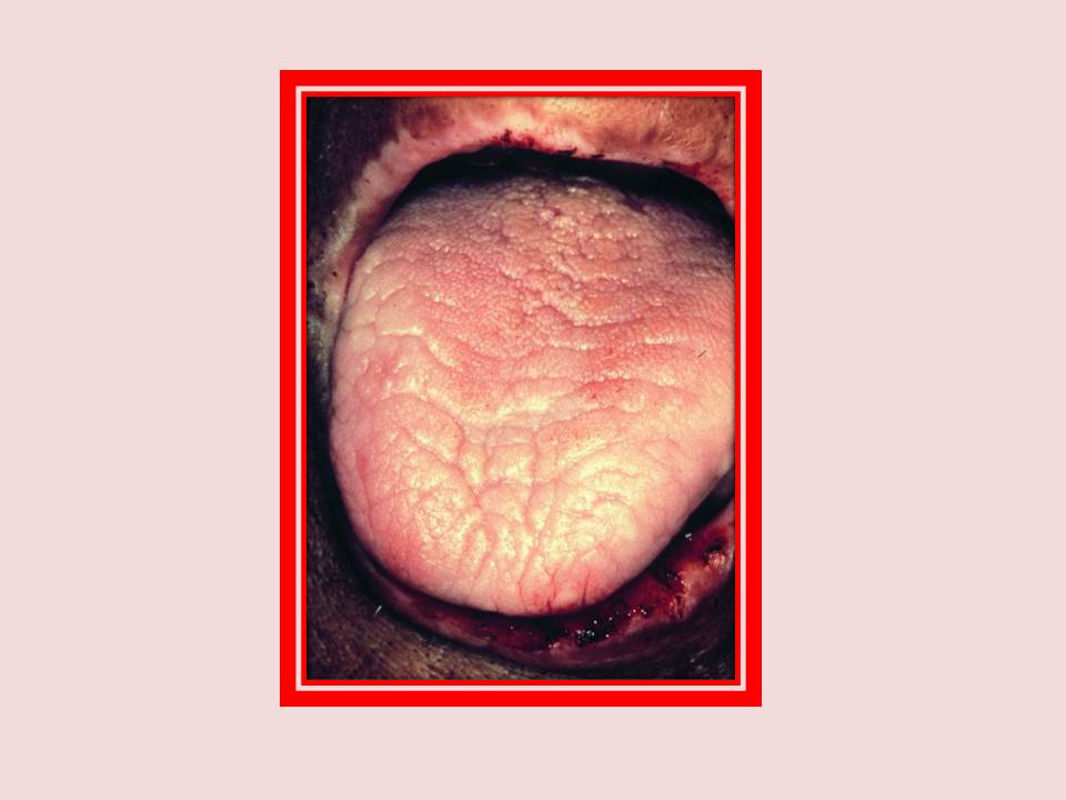
La dysphagie ferriprive
La dysphagie ferriprive. L'anémie due à une carence en fer avec la langue dépapillé, la dépigmentation de la lèvre supérieure et la érosion épithéliale de la lèvre inférieure.
|
|
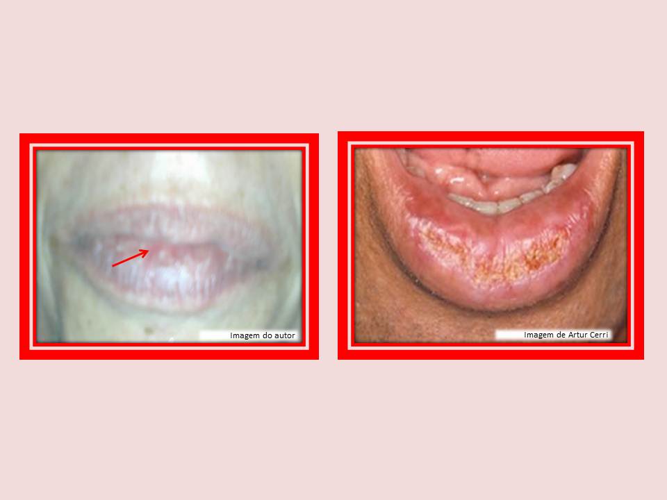
Chéilite actinique
Chéilite actinique. Ces sont des kératoses actiniques qu´ ont une incidence sur la lèvre vermillon. Se produisent surtout par l'exposition solaire.
|
|
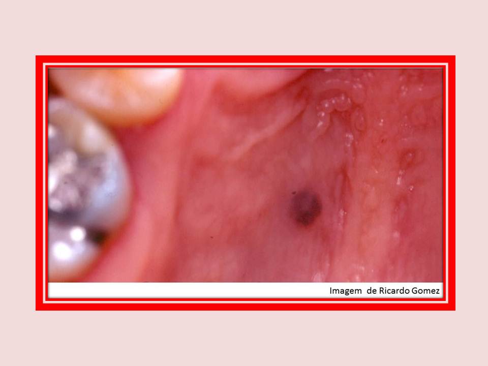
Nevo
Nevo. La présence d'une lésion pigmentée de la croissance récente, les bords irrégulièrs, de couleur variable, la présence d'une ulcération et des lésions satellites évoquent la possibilité d'un mélanome.
|
|
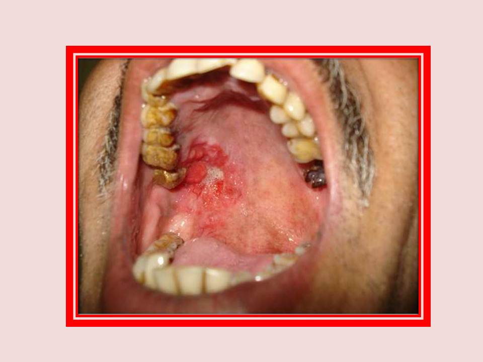
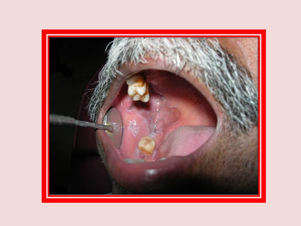
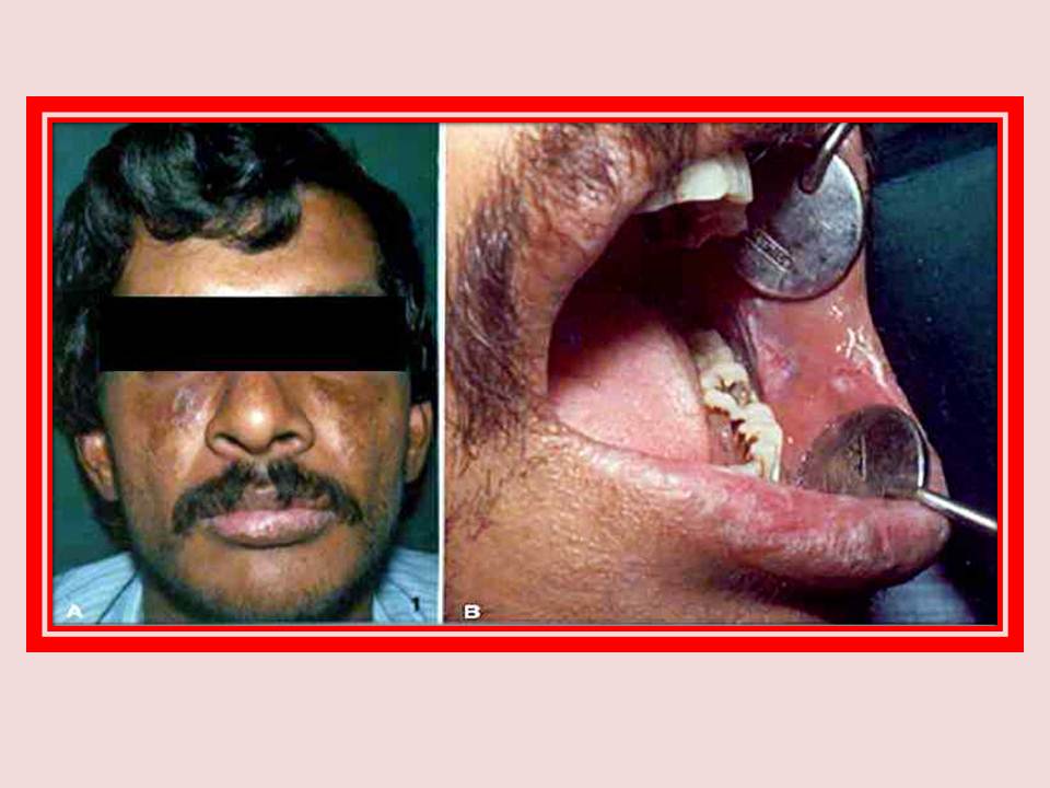
Fibrose sous-muqueuse orale
Fibrose sous-muqueuse orale avec une croissance maligne.
|
|
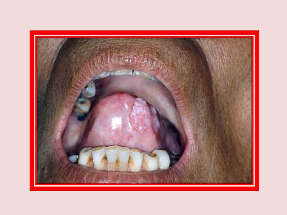
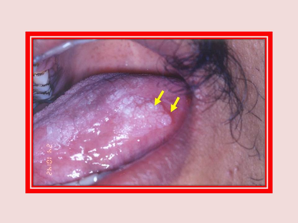
Leucoplasie
Leucoplasie homogène montrant la transformation maligne.
|
|
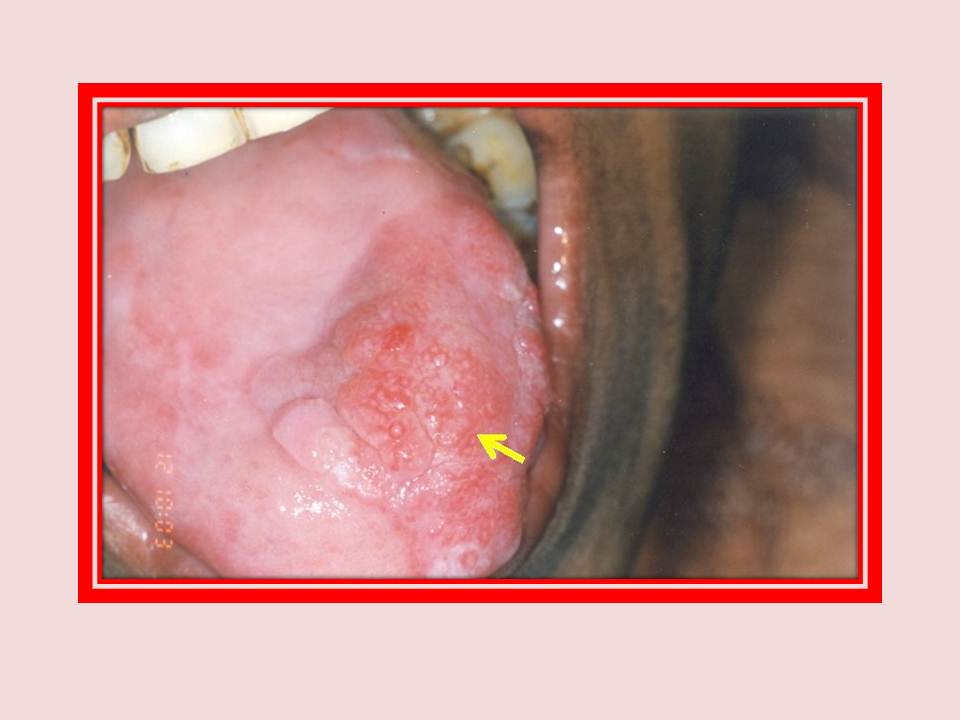
Fibrose sous-muqueuse orale
Fibrose sous-muqueuse orale avec une croissance maligne nodulaire proliférative sur la face dorsale antérieure de la langue.
|