|
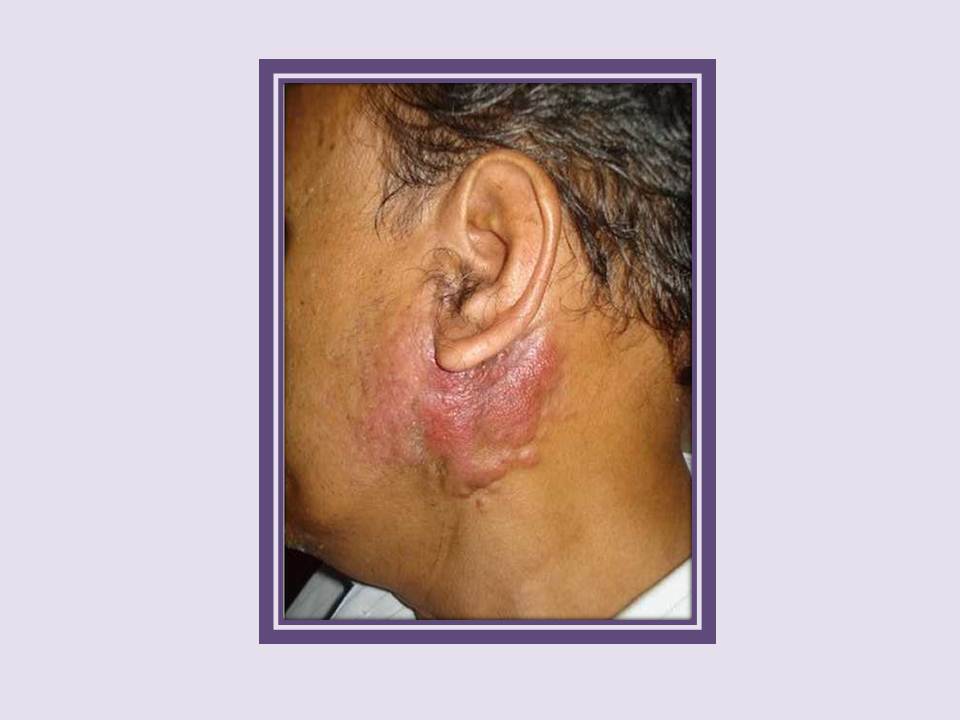
Carcinoma
Carcinoma of the left parotide gland infiltrating the overlying skin.
|
|
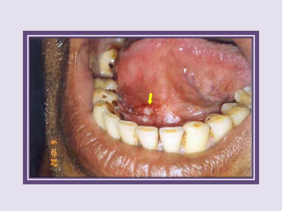
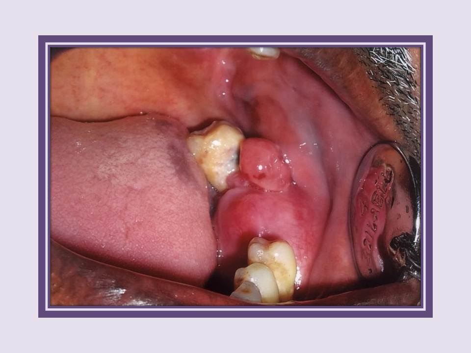
Carcinoma
Early invasive cancer in the floor of mouth. Note the erythematous ulcer proliferative growth in the floor of the mouth (arrow).
|
|
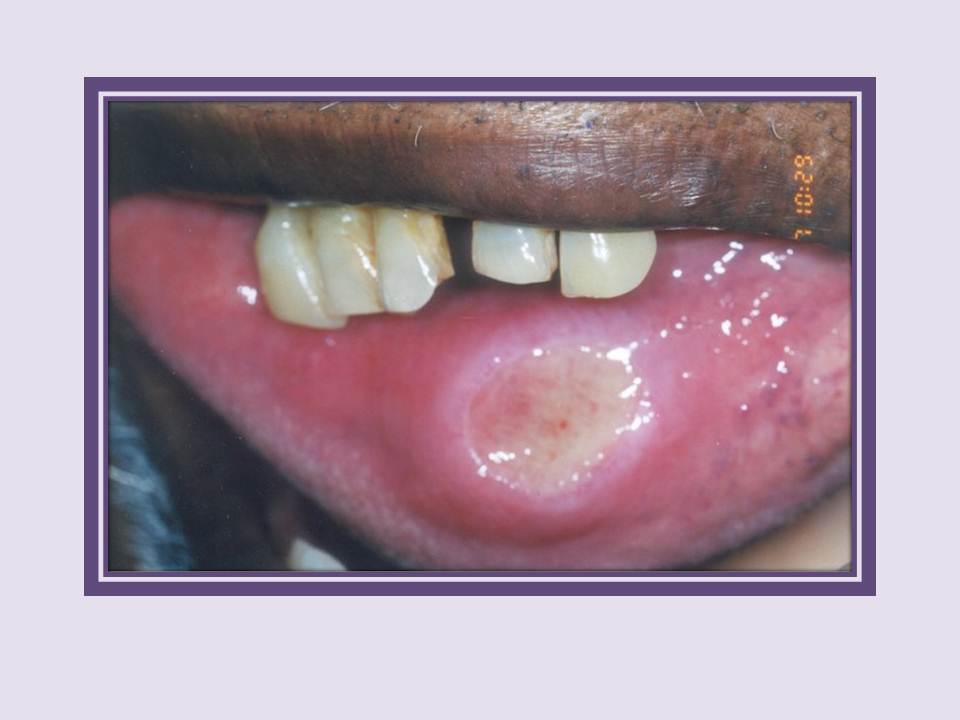
Carcinoma
Carcinoma of the lower lip. Note the punched out ulcerative lesion in the lower labial mucosa with raised and rolled out margins. The disease was caused by chronic irritation and trauma from the upper central incisors which have undergone attrition.
|
|
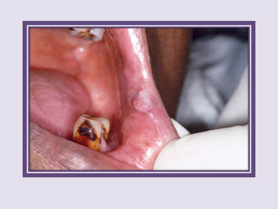
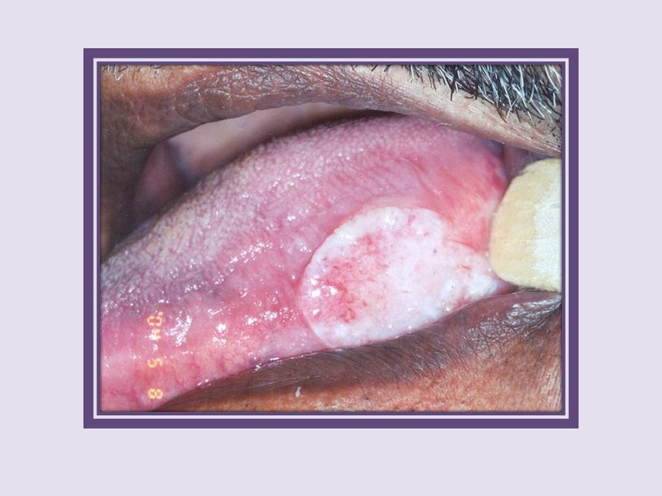
Carcinoma
Early carcinoma left commissure. An exophytic 1x1 cm growth in the left commissure.
|
|
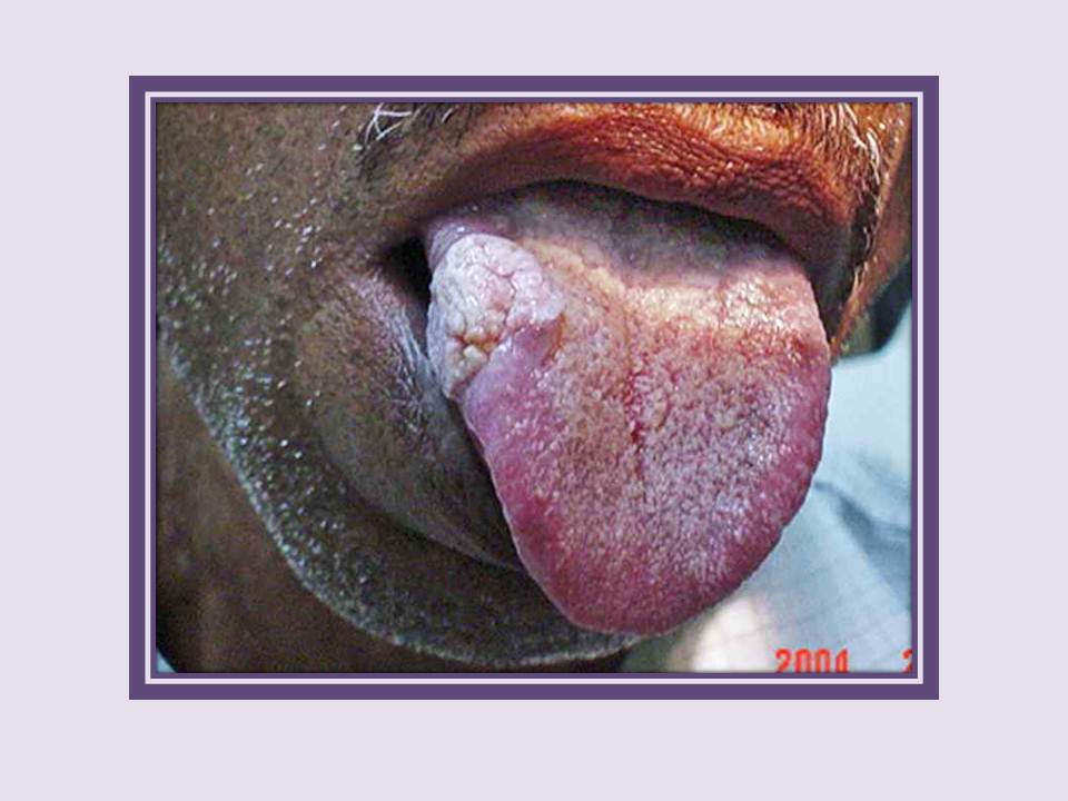
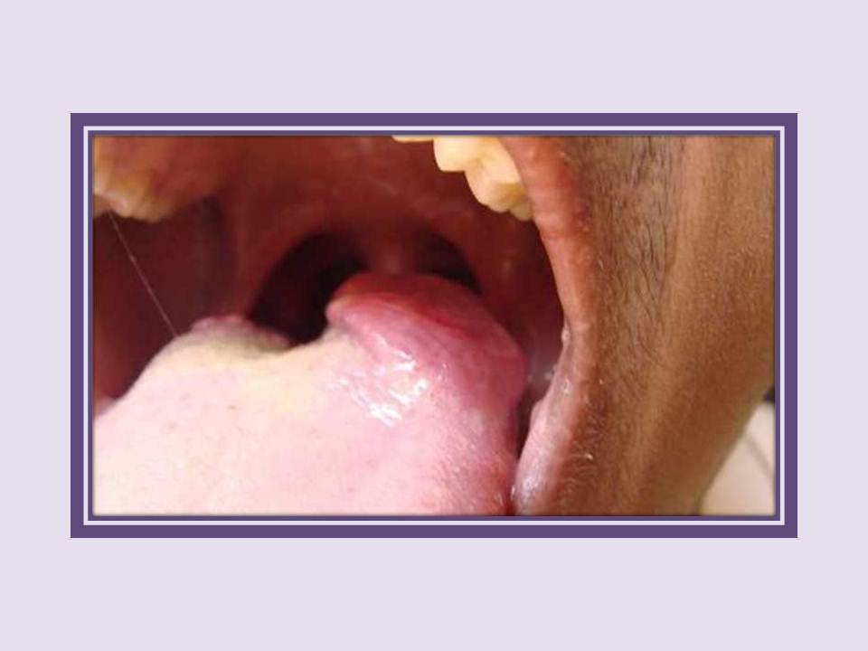
Carcinoma
Carcinoma of the tongue. Note the exophytic growth seen on the right lateral margin of tongue extending to the dorsum.
|
|
Carcinoma
Carcinoma of the tongue. Note the ulcer proliferative growth on the left lateral margin of the tongue.
|
|
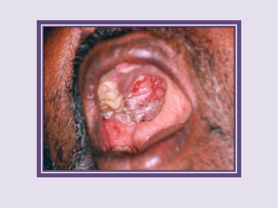
Carcinoma
Carcinoma on the right side of the hard palate. An exophytic growth of 5x4 cm extending from the right maxillary alveolus to the adjacent hard palate.
|
|
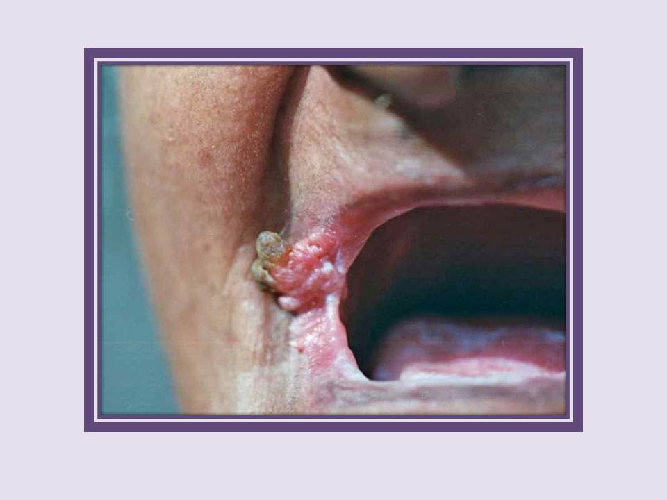
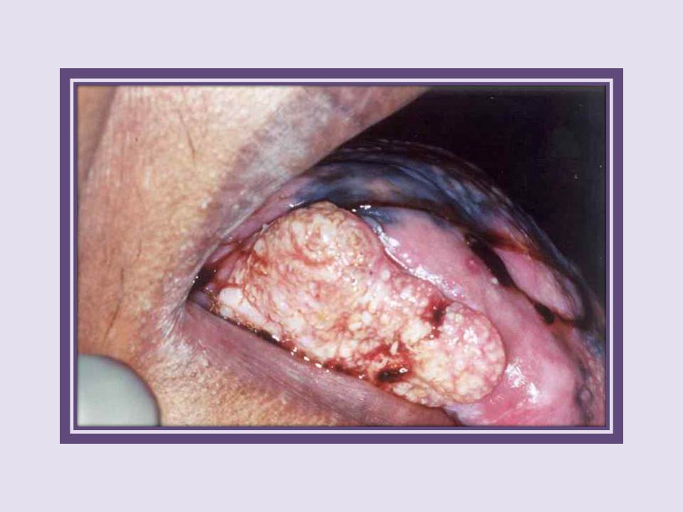
Carcinoma
Verrucous carcinoma right commissure. Note the exophytic growth with finger-like projections (arrows), extending to the right cheek in a patient with oral submucous fibrosis.
|
|
Carcinoma
Carcinoma of the tongue. Note the extensive ulceroproliferative growth on the right lateral margin of tongue.
|
|
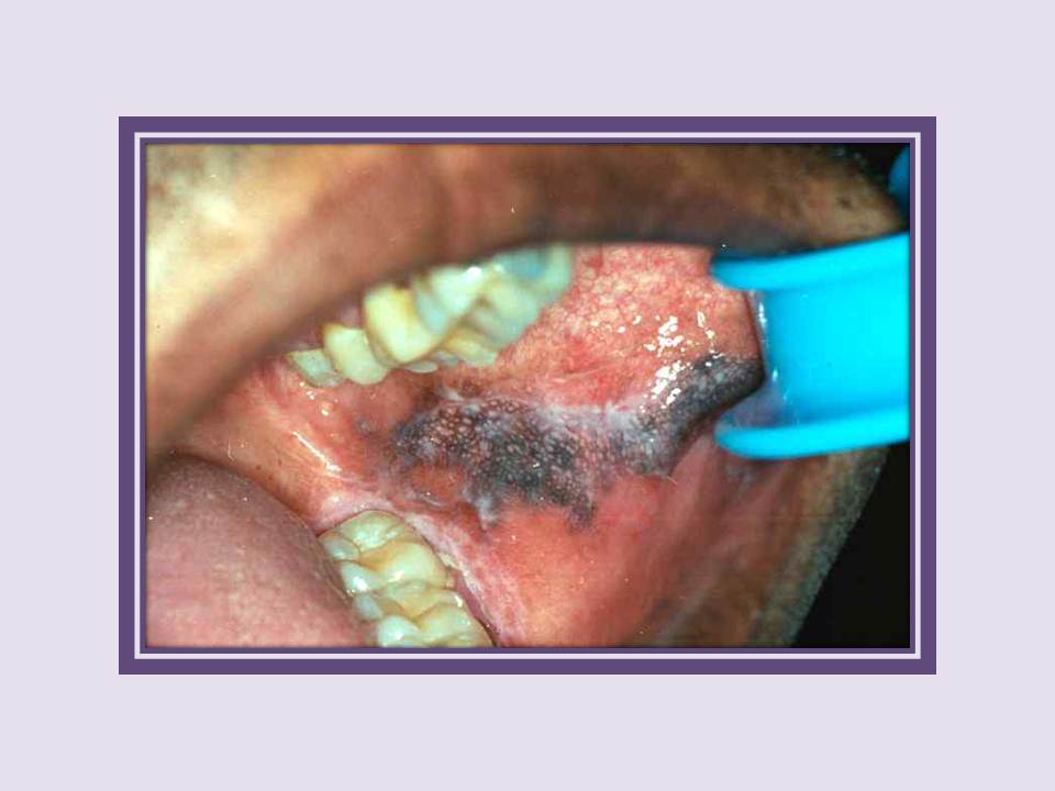
Melanoma
Malignant melanoma of the left buccal mucosa. Note the pigmented patch with nodular areas on the buccal mucosa of this 26-year-old man.
|
|
Lymphoma
Non-Hodgkin lymphoma of the oral cavity. Note the fleshy growth involving the right upper gingiva extending from the canine to the third molar.
|
|
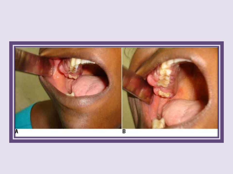
Lymphoma
Lymphoblastic lymphoma in a 12-year-old boy. Note the fleshy growth involving the left side of the posterior aspect of the tongue.
|
|
Metastatic tumor
Metastatic tumor in the left lower alveolus. Note the diffuse swelling in the left molar region with a fleshy 2x1 cm pedunculated mass near the last molar. The first two molars are missing.
|