|
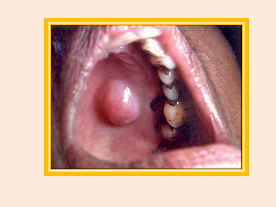
Adenoma
Adenoma pleomórfico. Observe o crescimento de 2x2 centímetros, macio e circunscrito no palato. A biópsia excisional revelou adenoma pleomórfico.
|
|
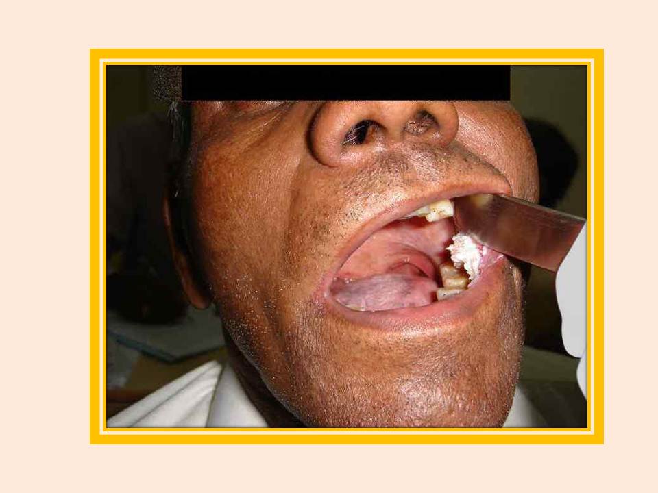
Papiloma
Papiloma. Um crescimento com projeções semelhantes a dedinhos.
|
|
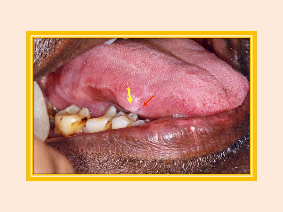
Fibroma
Fibroma, com ulceração superficial. Observe o volume (seta amarela) na margem lateral direita da língua, com uma úlcera linear (seta vermelha), devido à irritação crônica provocada pelo atrito com os dentes.
|
|
Pólipo
Pólipo fibroepitelial no dorso da língua. Um crescimento de 1x1 centímetros no lado direito do dorso da língua.
|
|
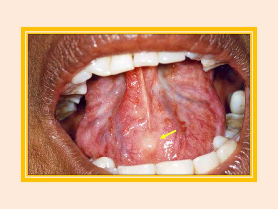
Lipoma
Lipoma no assoalho da boca. Observe o volume suave e amarelado na parte anterior do assoalho da boca, perto do freio lingual.
|
|
Granuloma
Tumor de células granulares. Observe o volume recoberto por epitélio normal.
|
|
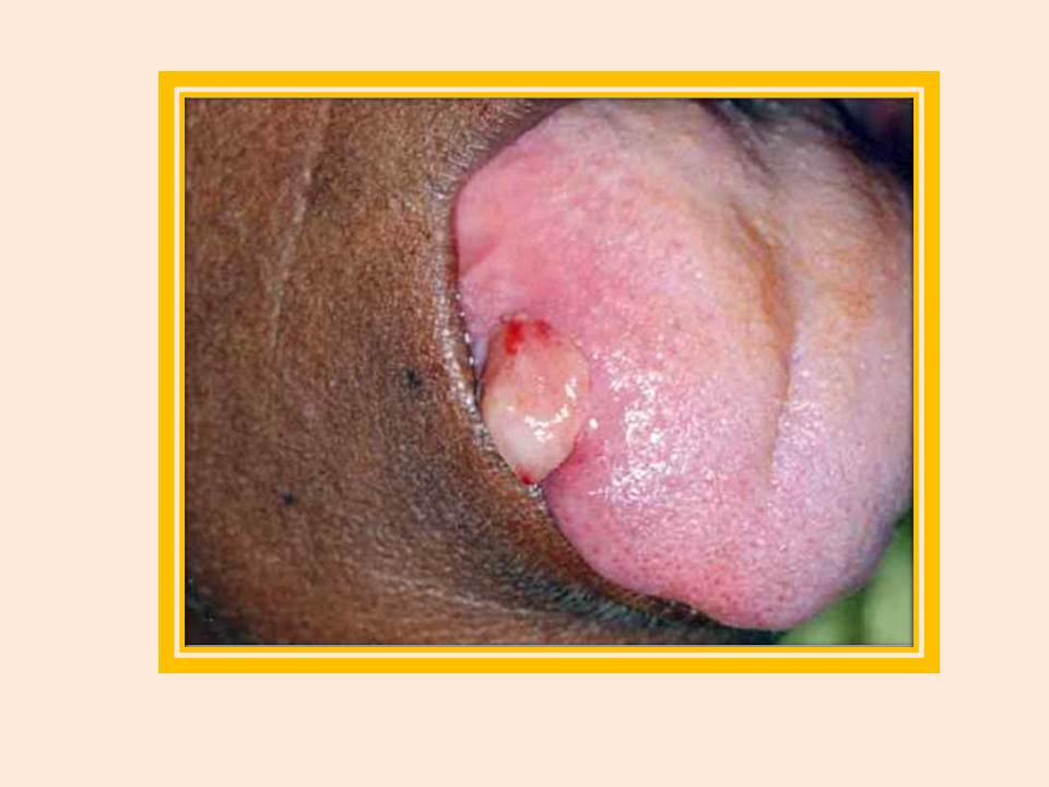
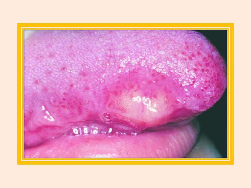
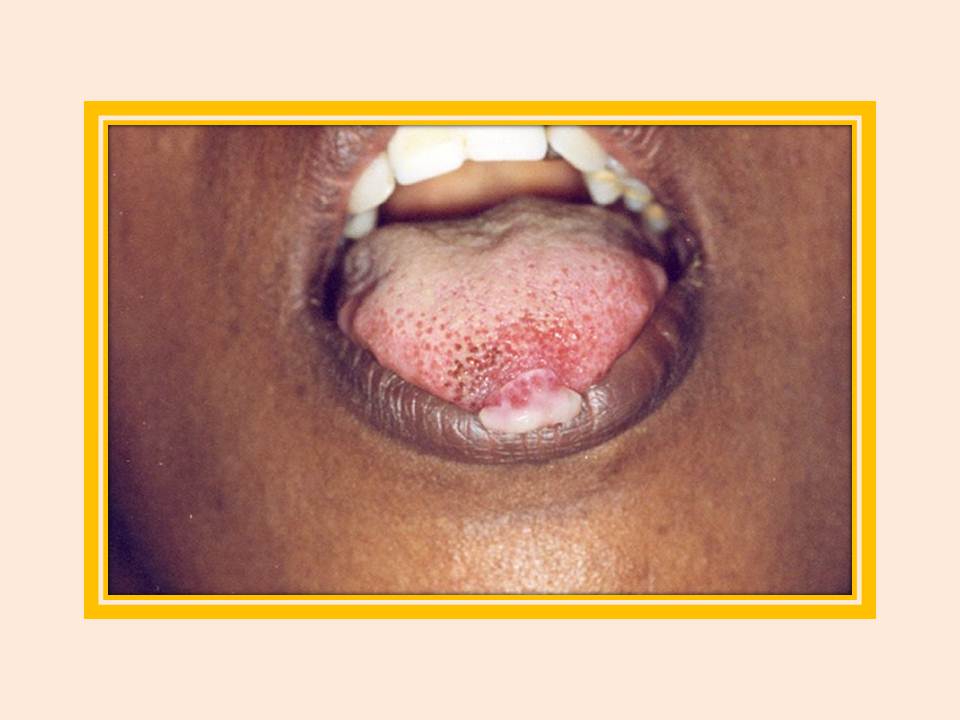
Hemangioma
Observe um crescimento de 0.5x0.5 centímetros no dorso da língua. Uma biópsia excisional revelou hemangioma ulcerado.
|
|
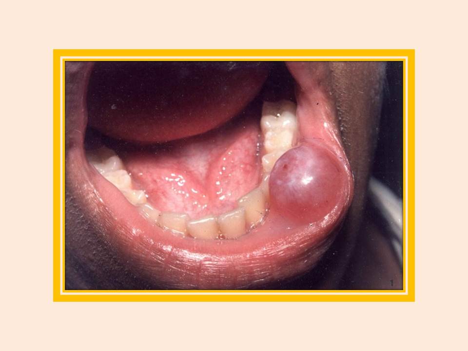
Hemangioma
Hemangioma no lábio inferior.
|
|
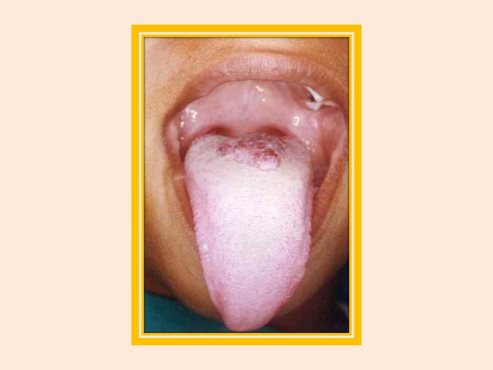
Hemangioendotelioma
Observe um crescimento de 1,5 X de 1,0 cm na ponta da língua. Um hemangioendotelioma ulcerado foi confirmado em biópsia excisional.
|
|
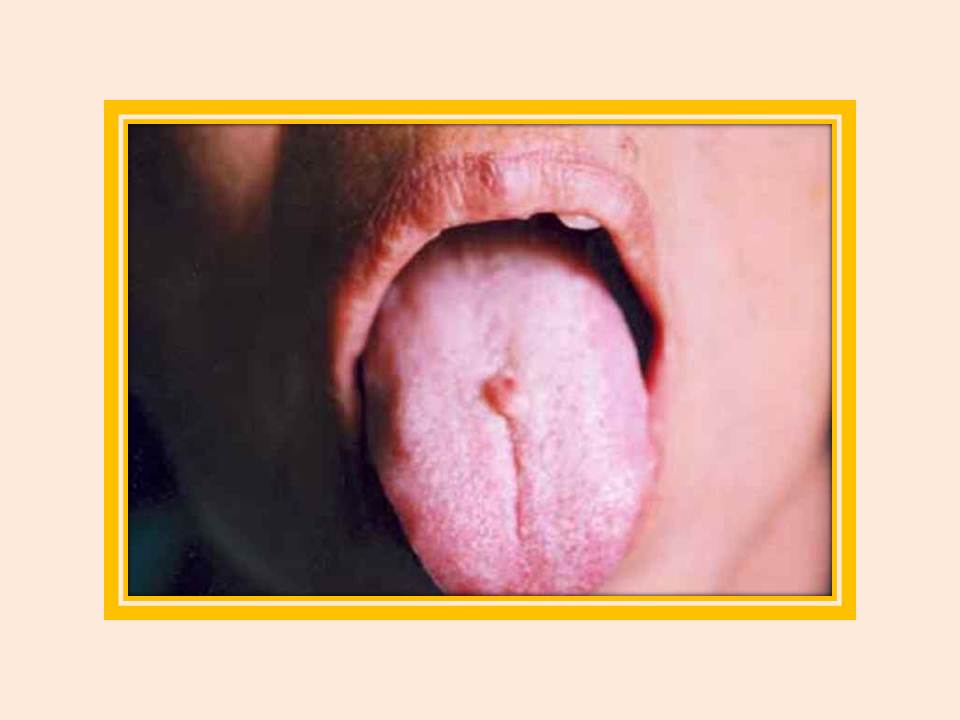
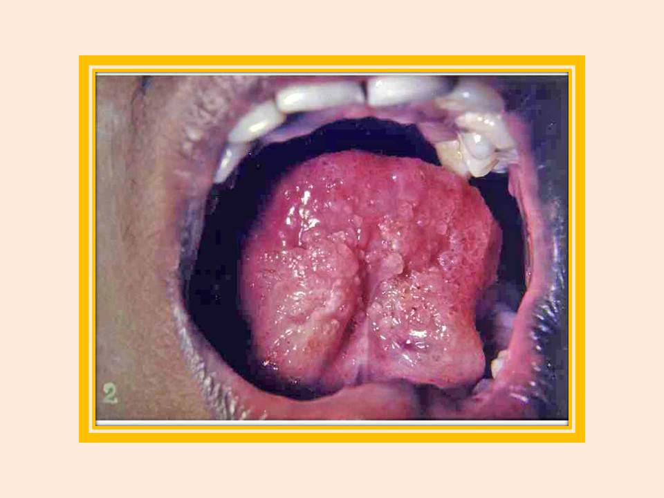
Linfangioma
Linfangioma na língua. Note-se a aparência proeminente com uma ranhura central, envolvendo a superfície ventral da língua.
|
|
Linfangioma
Linfangioma na língua.
|
|
Neurofibroma
Neurofibroma. Um crescimento bem definido, indolor, na gengiva.
|
|
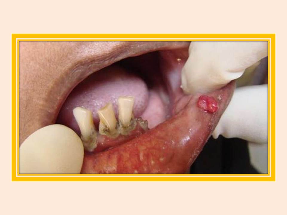
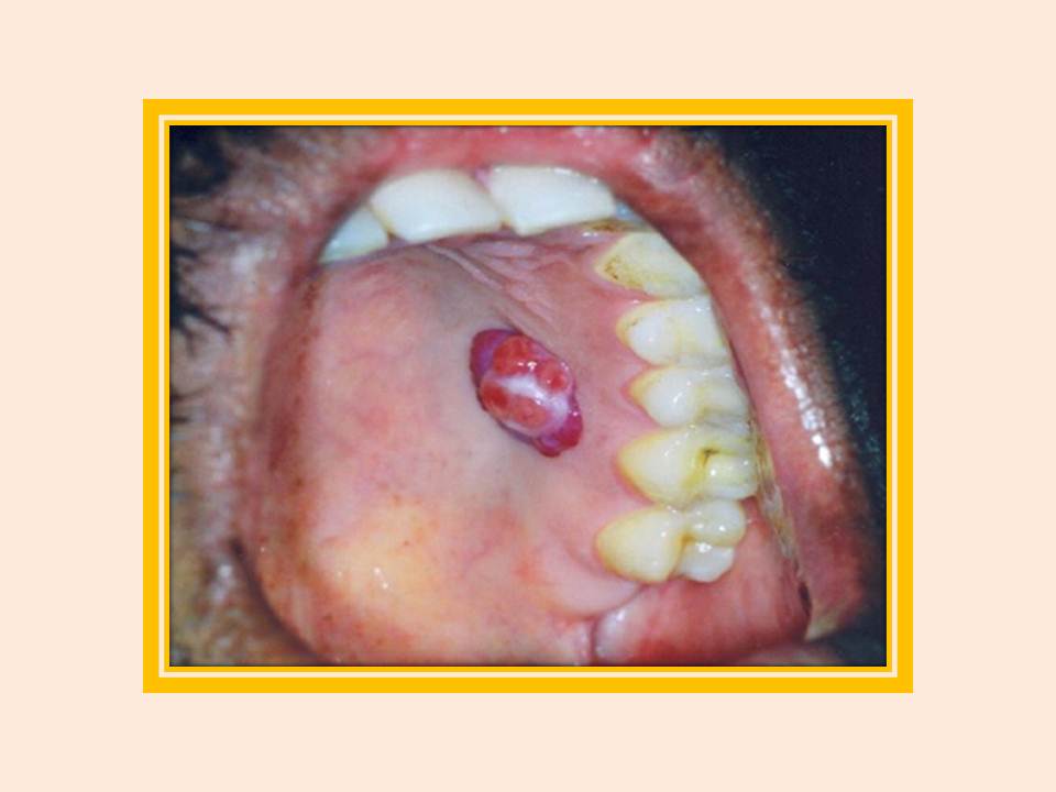
Granuloma
Granuloma piogênico. Observe o crescimento de 1.5x1.5 centímetros, vascularizado, no lado esquerdo do palato duro.
|
|
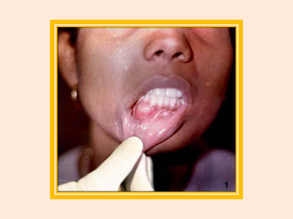
Mucocele
Mucocele. Note uma formação cística, firme, no lábio inferior.
|
|
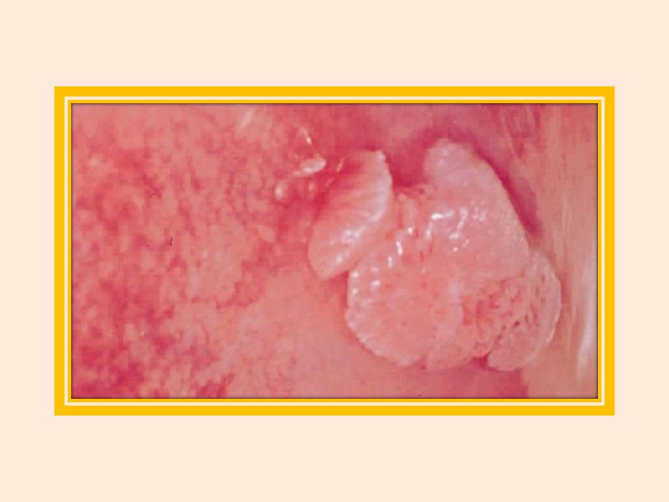
Condiloma acuminado
Condiloma acuminado. Crescimentos diversos, com aspecto de couve-flor.
|